5-in-1 ophthalmic diagnostic solution with speeds of up to 80,000 scans per second and high-quality real-color imaging.
OCTavius combines five key diagnostic functions — 3D OCT, fundus camera, angiography, biometry, and topography — into a single compact device. The system ensures seamless operation and delivers precise results necessary for making reliable clinical decisions.
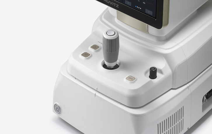
Thanks to the built-in computer, OCTavius eliminates the need for an external computer setup, ensuring maximum compactness. This allows for optimal use of space in the examination room and contributes to a more efficient workflow.
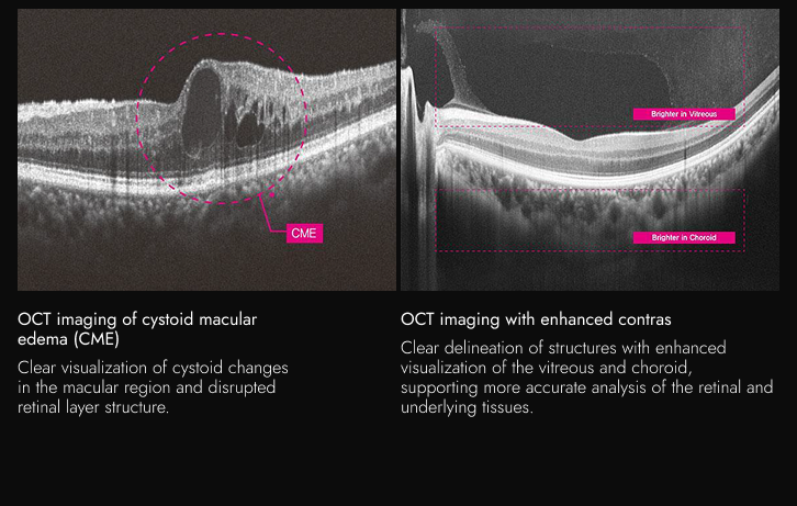
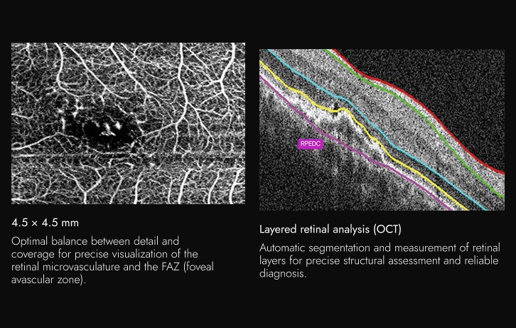
Even with involuntary eye movements or blinking, OCTavius provides stabilized 3D images with high resolution, thanks to scanning speeds of up to 80,000 A-scans per second. This ensures clear images without the need for retakes and minimizes the impact of patient movements. Precise visualization of the retinal layers allows detailed analysis of anatomical structures, including layer thickness and changes in the macular region, supporting more accurate diagnosis and confident clinical decision-making.
OCTavius provides true-color fundus imaging with precise contrast control in four levels, ensuring clear visualization of both the dark retinal areas and the bright regions around the optic disc. This allows detailed differentiation of arteries, veins, and fine microvasculature (capillaries), while automatic stitching of up to 7 images creates a wide panoramic view for fast and reliable clinical assessment.
OCTavius offers various scan sizes (3×3 to 9×9 mm) for precise visualization according to the location and extent of the lesion. With a single fast, non-contrast OCT-A scan, detailed and wide coverage of the retinal microvasculature is achieved, enhancing diagnostic accuracy and optimizing workflow.
OCT topography provides simultaneous 3D measurement of the anterior and posterior corneal surfaces with 16 types of corneal maps for precise assessment of thickness, curvature, and elevation. Optical biometry visualizes the axial structure from the cornea to the macula in a high-resolution 2D image, supporting accurate selection of the intraocular lens (IOL) based on the individual eye profile.
The integrated analysis enables fast and comprehensive assessment of each patient’s individual condition, including monitoring progression, comparison before and after treatment, and analysis against normative data.
Compatibility with Huvitz HIIS‑1 Cloud (optional) provides a web-based telemedicine platform that enables remote access, centralized data collection and storage, as well as advanced analyses—progression tracking, comparisons, and 3D visualization. This supports effective collaboration and informed clinical decision-making.